
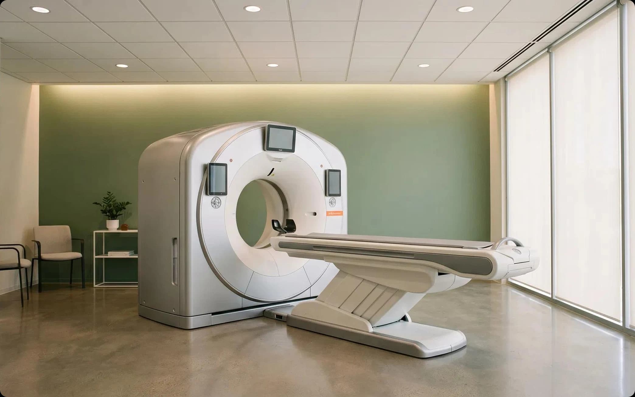
Preparation for FESS includes two key diagnostic components (nasal endoscopy and CT of the paranasal sinuses) and a set of pre-operative measures (medical preparation, management of antithrombotic therapy, patient information, anesthesia planning). Each stage is described below.
I. Nasal endoscopy — pre-operative assessment
Aims. Nasal endoscopy is the primary confirmatory test for CRS and a mandatory part of the pre-operative assessment. It shows inflammatory changes, polyps and anatomical anomalies and the nature of the discharge, which complements the CT findings and influences the surgical plan. [1][2][3]
Three-pass endoscopy technique. According to the AAO-HNS recommendations, a systematic endoscopic examination is performed in 3 passes: [1]
- First (lower) pass — visualization of the floor of the nasal cavity, the inferior turbinate/meatus, the Eustachian tube opening and the posterior wall of the nasopharynx
- Second (middle) pass — assessment of the middle turbinate, the olfactory cleft, the sphenoethmoidal recess, the superior turbinate and sometimes the sphenoid sinus ostium
- Third (upper) pass — examination of the nasal vault, the attachment of the middle turbinate to the lateral wall, the ostiomeatal complex, the uncinate process and the anterior ethmoid bulla
Preparing the nasal cavity for endoscopy. Topical vasoconstrictors and anesthetics are applied before the examination (unless contraindicated). The randomized study by Mishra et al. (2013, n=100) showed that packing with 4% lidocaine and xylometazoline gives better visualization and less discomfort and pain than a 10% lidocaine spray with xylometazoline, although it takes longer to prepare. An examination before and after decongestants is recommended to assess whether the swelling is reversible. [1][4]
Effect on the surgical plan. The study by Gardiner et al. (2023, n=346) showed that in 23.7% of patients referred for surgery for nasal obstruction, rigid endoscopy revealed findings not visible on anterior rhinoscopy. Endoscopic findings led to additional pre-operative tests in 14.5% and a change in the surgical plan in 7.5% of patients. Unexpected findings were most common in patients with previous nasal surgery and allergic rhinitis. [3]
Vining et al. (1993) found that in 9% of patients with sinusitis symptoms, endoscopy revealed pathology when the CT was normal (septal deviation, swelling of the middle meatus, enlarged adenoids), confirming that endoscopy and CT are complementary rather than interchangeable methods. [2]
II. Sinus CT — pre-operative protocol
Imaging standard. CT remains the gold standard for planning FESS. The AAO-HNS (2025) recommends: [5]
- Multidetector CT without contrast — the ACR-endorsed standard
- Thin-slice protocol: ideally ≤1 mm (GDG consensus), with 50% slice overlap to reduce artifacts on multiplanar reconstructions
- Three-plane reconstructions (axial, coronal, sagittal) — mandatory for assessing the anatomy and the extent of disease
- Contrast is not required for inflammatory disease; MRI complements CT when fungal sinusitis, a tumor or involvement of the orbit/skull base is suspected [6]
- Low-dose protocols (ALARA) are available and recommended, especially when repeat scans are needed
The CLOSE mnemonic — systematic CT review before FESS
O'Brien et al. (2016) proposed the CLOSE mnemonic for systematically assessing anatomical variants that predispose to surgical complications: [6][7]
| Letter | Structure | Clinical significance |
|---|---|---|
| C — Cribriform plate | Depth of the olfactory fossa (Keros classification I–III), asymmetry | Keros III (≥7 mm) — high risk of CSF leak and intracranial injury |
| L — Lamina papyracea | Dehiscence, thinning, medial bulging | Risk of orbital injury (hematoma, damage to the medial rectus muscle) |
| O — Onodi cell | A posterior ethmoid cell adjacent to the optic nerve/ICA | Risk of damage to the optic nerve and internal carotid artery |
| S — Sphenoid sinus (pneumatization) | Type of pneumatization (conchal, presellar, sellar), relation to the ICA | Lateral pneumatization → dehiscence of the ICA wall → catastrophic bleeding |
| E — Ethmoidal artery (anterior) | Position (in the skull base vs. hanging free in the cavity), dehiscence of the canal | Free-hanging artery → risk of retraction if injured → retrobulbar hematoma |
III. Pre-operative medical preparation
Systemic corticosteroids in CRSwNP. Pre-operative systemic corticosteroids in CRS with polyps reduce the polyp burden, improve visualization of the surgical field and reduce intraoperative blood loss. The systematic review by Chrysouli et al. (2022, 15 studies) confirmed a significant effect of corticosteroids on polyp size, symptoms and markers of inflammation. EPOS recommends the use of nasal corticosteroids before FESS. The AAO-HNS (2025) notes that a short course of oral corticosteroids may be considered perioperatively in CRSwNP, although the optimal dose and duration are not standardized. [8][9]
The RCT by Kominsky et al. (2022, n=23) compared low, medium and high doses of prednisolone before FESS: the medium dose showed a trend towards better visualization than the low dose, but no statistically significant differences between the groups were found. [10]
Management of antithrombotic therapy. Anticoagulants and antiplatelet agents are routinely stopped before FESS to reduce the risk of perioperative bleeding. The systematic review by Boyi et al. (2024) found no clear recommendations on when to resume antithrombotic therapy after FESS — recommendations in the literature are contradictory. The decision is made individually, taking into account the patient’s thrombotic risk. [11]
For patients who cannot stop antithrombotic therapy, in-office balloon ostial dilation may be a safe alternative to FESS (Higgins et al., 2020, n=35). [12]
IV. Anesthesia planning
FESS is usually performed under general anesthesia, although office procedures under local anesthesia are becoming more common. The narrative review by Stamenkovic et al. (2023) summarized best practices in perioperative care: [13][14]
- Topical vasoconstrictors (xylometazoline, oxymetazoline, adrenaline on pledgets) — mandatory to optimize the surgical field
- Positioning — reverse Trendelenburg (15–30°) to reduce venous pressure
- Controlled hypotension — moderate hypotension improves visualization
- TIVA vs. inhalation anesthesia — 4 of 5 meta-analyses and 6 of 11 RCTs showed an advantage of TIVA for visualization of the surgical field, but the effect depends more on adjuvant drugs (remifentanil, α2-agonists) than on the choice of the main anesthetic technique
- It is recommended to use the technique the anesthesiologist is most comfortable with
V. Image-guided surgery (IGS)
The AAO-HNS endorses image-guided surgery (IGS) for the following indications: [15][16]
- Revision sinus surgery
- Distorted anatomy (congenital, post-traumatic, postoperative)
- Extensive nasal polyposis
- Disease of the frontal, posterior ethmoid and sphenoid sinuses
- Benign and malignant sinonasal tumors
The narrative review by Beswick & Ramakrishnan (JAMA Otolaryngology, 2020) showed that there is no convincing evidence that IGS improves surgical outcomes (completeness of dissection, QOL, revision rates), but level 2A data from systematic reviews suggest a possible reduction in overall and major surgical complications in complex cases. The decision to use IGS remains with the surgeon, based on the complexity of the case and personal experience. [17]
When IGS is planned, the CT must be performed according to a specific navigation protocol (thin slices, a defined data format). [5][18]
VI. Patient information (pre-operative counseling)
The AAO-HNS (2025) recommends that the surgeon educate the patient before surgery on the following: [5]
- CRS is a chronic disease; surgery is not a “cure” but part of a comprehensive plan
- Expected postoperative care: pain control, debridement, medical therapy (nasal irrigation, topical corticosteroids), activity restrictions, time off work
- The frequency and duration of postoperative visits
- The potential need for revision surgery (20% within 5 years in CRSwNP)
- Risks and complications: bleeding, infection, CSF leak, orbital injury, smell disorders
Summary table: stages of preparation for FESS
| Stage | Components | Key recommendations |
|---|---|---|
| Nasal endoscopy | 3-pass technique, topical anesthesia + decongestant | Mandatory; reveals pathology not visible on CT in 9% of patients; changes the plan in 7.5% |
| Sinus CT | Multidetector, no contrast, ≤1 mm, 3 planes | Gold standard; CLOSE review; an up-to-date CT is mandatory before revision |
| Medical preparation | Systemic corticosteroids (in CRSwNP), stopping anticoagulants | Corticosteroids reduce polyps and blood loss; the optimal dose is not standardized |
| Anesthesia planning | TIVA/inhalation, controlled hypotension, positioning | TIVA may improve visualization; adjuvants matter more than the choice of technique |
| Navigation (IGS) | Electromagnetic/optical | Indicated for revision, distorted anatomy, extensive polyposis |
| Patient information | Risks, postoperative care, chronic nature of the disease | 3D reconstructions improve understanding |
Sources
- 1.Rhinosinusitis: Developing Guidance for Clinical Trials — J Allergy Clin Immunol, 2006
- 2.The Importance of Preoperative Nasal Endoscopy in Patients With Sinonasal Disease — The Laryngoscope, 1993
- 3.The Role of Nasal Endoscopy in the Preoperative Evaluation of Nasal Airway Obstruction — Otolaryngology–Head and Neck Surgery, 2023
- 4.Preparation of Nose for Nasal Endoscopy: Cotton Pledget Packing Versus Topical Spray — Eur Arch Otorhinolaryngol, 2013
- 5.Clinical Practice Guideline: Surgical Management of Chronic Rhinosinusitis (AAO-HNS) — Otolaryngology–Head and Neck Surgery, 2025
- 6.ACR Appropriateness Criteria Sinonasal Disease: 2021 Update — JACR, 2022
- 7.The Preoperative Sinus CT: Avoiding a «CLOSE» Call With Surgical Complications — Radiology, 2016
- 8.The Effect of the Premedication With Systemic Corticosteroids and Antibiotics on Inflammation and Intraoperative Bleeding During FESS for CRSwNP — J Craniofac Surg, 2022
- 9.Clinical Practice Guideline: Adult Sinusitis Update — Otolaryngology–Head and Neck Surgery, 2025
- 10.Seeking an Optimal Dose of Preoperative Corticosteroids in Chronic Rhinosinusitis With Nasal Polyposis: A Randomized Controlled Trial (Kominsky et al.) — American Journal of Otolaryngology, 2022
- 11.Restarting Antithrombotic Therapies After Endoscopic Sinus Surgery: A Systematic Review (Boyi et al.) — Ann Otol Rhinol Laryngol, 2024
- 12.In-Office Balloon Sinus Ostial Dilation With Concurrent Antiplatelet and Anticoagulant Therapy for Chronic Rhinosinusitis Without Nasal Polyps (Higgins et al.) — Ann Otol Rhinol Laryngol, 2020
- 13.Chronic Rhinosinusitis with Nasal Polyps (Hopkins C.) — NEJM, 2019
- 14.Perioperative Management and Surgical Field Optimization in Functional Endoscopic Sinus Surgery (Stamenkovic et al.) — Minerva Anestesiologica, 2023
- 15.Image-Guided Surgery in Otolaryngology: A Review of Current Applications and Future Directions in Head and Neck Surgery (Bessen et al.) — Head & Neck, 2021
- 16.Surgical Navigation in the Anterior Skull Base Using 3-Dimensional Endoscopy and Surface Reconstruction — JAMA Otolaryngology–Head & Neck Surgery, 2024
- 17.The Utility of Image Guidance in Endoscopic Sinus Surgery: A Narrative Review (Beswick & Ramakrishnan) — JAMA Otolaryngology–Head & Neck Surgery, 2020
- 18.Clinical Consensus Statement: Appropriate Use of Computed Tomography for Paranasal Sinus Disease — Otolaryngology–Head and Neck Surgery, 2012
- 19.Allegations of Failure to Obtain Informed Consent in Otolaryngology: Evidence-Based Recommendations for Sinus Surgeons — American Journal of Rhinology & Allergy, 2023
- 20.«When Patients See What the Surgeon Sees»: 3D Imaging Creates New Understanding in Preoperative Counseling for Endoscopic Sinus Surgery — Acta Oto-Laryngologica, 2026
- 21.Comorbid Chronic Rhinosinusitis and Asthma: Shared Risk Factors and Treatment Implications — an EAACI Task Force Report — Allergy, 2026
- 22.Aspirin-Exacerbated Respiratory Disease — NEJM, 2018
- 23.Systemic Predictors of Eosinophilic Chronic Rhinosinusitis — American Journal of Rhinology & Allergy, 2018
- 24.Systemic Biomarkers of Eosinophilic Chronic Rhinosinusitis — Current Opinion in Allergy and Clinical Immunology, 2020
Still have questions?
Book a consultation with a specialist
Book